Newsletter Subscribe
Enter your email address below and subscribe to our newsletter

 ByJohn A
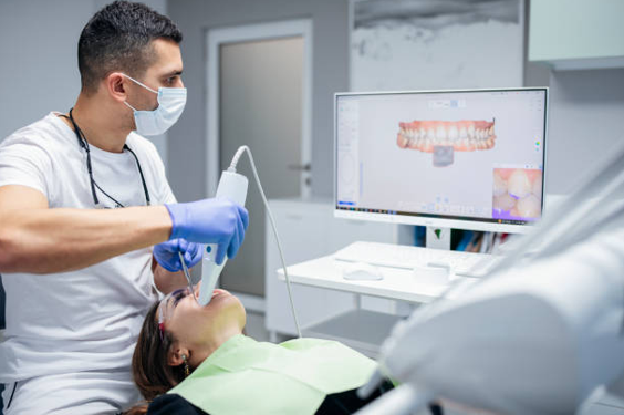
Three-dimensional dental scans help implant providers evaluate the jawbone, locate important nerves, examine sinus anatomy, and determine suitable positions for dental implants. Cone-beam computed tomography (CBCT) produces detailed images of oral structures that conventional dental X-rays may not show adequately. Combined with a clinical examination, these images help the dental team assess bone availability, identify anatomical concerns, and plan implant placement with greater precision. For patients considering dental implants Tysons Corner VA, this evaluation provides essential information before surgery. G4 Dental Implants uses the implant planning process to help patients understand their oral anatomy and the factors that influence treatment decisions.
A scan is only one part of the assessment. It cannot independently determine gum health, confirm every aspect of bone quality, or guarantee a successful result. The treating clinician must interpret the images alongside the patient’s medical and dental history, examination findings, and restorative goals.
Cone-beam computed tomography is an imaging technique that creates three-dimensional views of the teeth, jawbone, and surrounding anatomical structures. Unlike a conventional two-dimensional dental X-ray, which compresses structures into a flat image, CBCT allows the clinician to examine the jaw from different angles and assess its width, height, and shape.
During a CBCT scan, a machine rotates around the patient’s head and captures multiple images. Software combines these images into a three-dimensional representation of the area being examined. The scan typically takes only a short period to capture, although the overall appointment may include additional assessment and planning.
The clinician selects the appropriate scan area based on the treatment being considered. Imaging should follow the principle of using the smallest field of view and lowest radiation exposure that provide the information needed for safe clinical decision-making.
See also: What is Considered a Dental Emergency? (And When It Can Wait)
A dental implant must be supported by adequate bone. After a tooth is lost, the surrounding jawbone can gradually change in height and width. Periodontal disease, previous infection, trauma, and prolonged tooth loss may also affect the available bone.
3D dental implant imaging helps the clinician evaluate the dimensions of the proposed implant site before selecting an implant size or planning the surgical approach.
A CBCT scan can show the vertical height and horizontal width of the jawbone at the intended placement site. These measurements help identify areas where bone may be too narrow or where the available height is limited by nearby anatomical structures.
For example, a patient may have sufficient bone height but inadequate width to accommodate an implant safely. A two-dimensional X-ray may not reveal the full extent of this limitation because it does not show the jaw’s width directly. Three-dimensional images help the clinician assess the available space and determine whether the planned implant position is appropriate.
The jawbone is not a uniform block of tissue. Its contours can vary along the ridge, particularly after tooth extraction or bone loss. CBCT images can reveal irregularities, depressions, and defects that may influence implant positioning.
These findings help the clinician decide whether the site can accommodate an implant directly or requires additional evaluation for procedures such as bone grafting. The final decision also depends on the clinical examination, the patient’s healing capacity, and the desired position of the replacement tooth.
Jawbone density for implants affects how the implant interacts with the surrounding bone during placement and healing. CBCT images provide information about bone structure and may help clinicians estimate relative density. However, conventional CBCT grayscale values are not standardized enough to serve as a universally reliable measurement of bone mineral density.
A scan cannot guarantee how firmly an implant will hold or predict its long-term integration by itself. The clinician considers the imaging findings alongside bone anatomy, surgical observations, and other relevant patient factors.
Important anatomical structures sit close to some implant sites. Their positions can limit the available space and influence the safest direction and depth for implant placement.
In the lower jaw, the inferior alveolar nerve travels through a bony canal and supplies sensation to the lower teeth, lower lip, and chin. An implant placed too close to this nerve can cause sensory disturbances, including numbness, tingling, or altered sensation.
CBCT helps the clinician identify the canal’s location and measure the distance between the proposed implant site and the nerve pathway. This information supports decisions about implant length, angle, and placement depth. It also helps the clinician recognize anatomical variations that may not be clear on conventional X-rays.
Imaging reduces uncertainty, but it does not eliminate nerve injury risk. Accurate interpretation, appropriate surgical technique, and careful planning remain necessary.
The maxillary sinuses are air-filled spaces located above the upper back teeth. After tooth loss, the bone beneath a sinus may become thinner, leaving less vertical bone available for implant placement.
A CBCT scan shows the relationship between the sinus floor and the intended implant site. It may also reveal features such as sinus septa, thickened sinus lining, or other abnormalities that warrant further assessment.
These findings help the clinician decide whether an implant can be placed within the available bone or if a sinus augmentation procedure needs consideration. An imaging abnormality does not automatically mean surgery must be delayed or cancelled. Its clinical significance depends on the finding and the patient’s symptoms and history.
A three-dimensional image provides anatomical information, but it does not replace a full dental examination. Successful treatment planning requires an understanding of the patient’s oral health, general health, bite, and ability to maintain the implant.
The clinical evaluation commonly includes an assessment of gum health, remaining teeth, bite alignment, oral hygiene, and the condition of the tissues surrounding the proposed implant site. The dentist may also review previous dental procedures, medical conditions, medications, tobacco use, and a history of periodontal disease.
These factors matter because an implant can be positioned correctly in the bone yet face complications if the surrounding gums are unhealthy or the bite places excessive force on the restoration.
The clinician also evaluates the planned replacement tooth. Its shape, position, and relationship with adjacent teeth affect the implant’s location. Placing an implant based solely on the available bone may create difficulties with cleaning, appearance, or bite function.
At G4 Dental Implants, the planning discussion should connect the scan findings with the patient’s oral health and restorative objectives. Patients benefit from understanding the reason for the recommended implant position and any additional procedures that may be required.
Dental implant surgical planning combines three-dimensional imaging with clinical findings and the intended design of the final restoration. The clinician uses the available information to determine the proposed implant position, orientation, and dimensions.
The planning process generally involves:
Digital planning software may allow the clinician to position a virtual implant within the CBCT image. In selected cases, this information can be used to design a surgical guide that helps transfer the planned position to the patient’s mouth.
A surgical guide does not remove the need for professional judgment. Its accuracy depends on image quality, planning, fabrication, fit, and correct use during surgery. The clinician must also account for any differences between the planned conditions and the actual surgical site.
CBCT imaging can provide valuable information that may not be available from conventional dental X-rays. Its benefits include clearer assessment of jawbone dimensions, improved visualization of nerve pathways, evaluation of sinus anatomy, and better-informed implant positioning. These details can help clinicians identify anatomical limitations before surgery and explain treatment options more clearly.
However, CBCT uses ionizing radiation. Although the exposure from dental CBCT is generally lower than that from many medical CT examinations, it is typically higher than that from routine dental X-rays. The actual dose varies with the machine, scan settings, and field of view.
For this reason, CBCT should be prescribed when the additional information is expected to affect diagnosis or treatment planning. It is not necessary for every dental implant patient, and repeat scans should be justified by a clinical need.
The images may also show incidental findings outside the immediate implant site. Some findings require further evaluation, and the clinician must review the relevant image volume rather than focusing exclusively on the proposed implant location.
Patients should inform the dental team about relevant medical history and pregnancy status before imaging so the provider can determine the appropriate examination. The objective is to obtain sufficient diagnostic information while minimizing unnecessary radiation exposure.
CBCT findings sometimes reveal that the initial treatment plan needs modification. A narrow ridge, limited bone height, an unfavourable nerve position, or a sinus that extends close to the implant site may require a different implant position or additional treatment.
The clinician may also recommend treating active gum disease, addressing an infection, or evaluating a nearby tooth before proceeding. Some patients may benefit from bone grafting, while others may have enough bone for implant placement without augmentation.
Not every anatomical variation requires an additional procedure. The appropriate response depends on the available bone, the intended restoration, the patient’s health, and the risks associated with each option.
Patients should ask the provider to explain the findings in plain language, including which details are clearly visible on the scan, which conclusions rely on clinical examination, and what uncertainties remain. This discussion helps patients make informed decisions rather than assuming that a three-dimensional image automatically determines the treatment.
Before proceeding with treatment, patients can make the consultation more useful by bringing relevant dental records and sharing details about previous extractions, gum treatment, medical conditions, and current medications. Existing X-rays or scans may be helpful if they are recent, diagnostically adequate, and suitable for the proposed evaluation.
It is also useful to ask how the implant’s planned position will support the final tooth, what the scan reveals about nearby anatomical structures, and if any additional procedures may be necessary. Patients should understand the benefits and limitations of the recommended imaging method and the alternatives available.
A clear explanation of these findings helps establish realistic expectations. It also gives patients a better understanding of the relationship between bone anatomy, implant placement, restoration design, and long-term maintenance.
Three-dimensional imaging gives implant clinicians a detailed view of the jawbone and nearby structures, helping them assess the available bone, identify nerve and sinus locations, and plan implant placement with greater anatomical awareness. Its greatest value comes from combining scan findings with a thorough examination, careful restorative planning, and an understanding of the patient’s health.
G4 Dental Implants can help patients understand how imaging findings relate to their proposed treatment and what those findings mean for their individual circumstances. If you are considering implant treatment, fix an appointment for a consultation to discuss your oral health, the need for CBCT imaging, and the factors that may influence your treatment plan.
An informed decision begins with understanding the evidence, the limitations of the scan, and the clinical reasoning behind the recommended approach. That foundation helps patients participate confidently in decisions about restoring missing teeth and maintaining their oral health.